Anthony Holland’s RF-plasma experiments occupy an unusual place in the history of bioelectromagnetics. The work began far outside conventional oncology, reached a real university cancer laboratory, produced recorded and sometimes quantified effects in cultured human cancer cells, and then ran directly into the standard that separates an arresting observation from a proven treatment: controls, replication, peer review, and eventually clinical testing.
This is the story of what Holland and his collaborators actually built, what they reported seeing under the microscope, what later experiments measured—and what the evidence still does not establish.
Research context: Every result discussed here was produced in cell culture. Holland’s RF-plasma approach has not been established as safe or effective for treating cancer in people. Nothing in this article is medical advice, and none of the RF/plasma equipment sold by Sacred Geometry is a medical device.
From frequency synthesis to an enclosed-gas plasma antenna
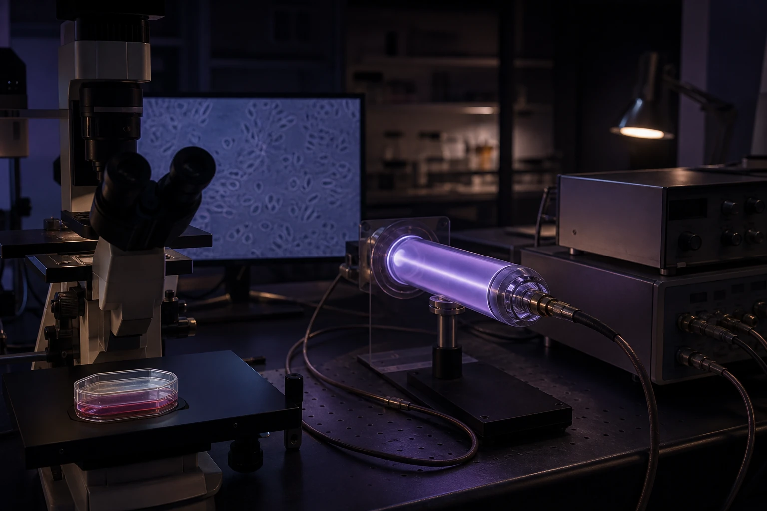
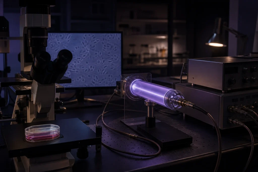
Holland was a longtime Skidmore College music professor, orchestra conductor, composer, and specialist in digital audio synthesis. His central technical idea was not that audible music would kill cancer. He used programmable waveforms to control an RF transmitter and power amplifier, which in turn energized a sealed, gas-filled glass tube. The glowing plasma tube acted as an antenna for an oscillating, pulsed electric field.
The later papers call this oscillating pulsed electric fields, or OPEF. A typical research chain consisted of a programmable frequency source, amplitude modulation, an RF carrier, a high-power RF amplifier, an impedance tuner, and an enclosed helium plasma tube positioned near a cell culture. The pulse-repetition or modulation frequency was varied while the RF carrier and plasma assembly delivered the field.
That distinction matters. This was not a loudspeaker experiment, and it was not simply “playing a frequency” into a room. It was an RF electronics platform whose visible plasma tube served as the radiating element.
A genuine university-laboratory test
Holland’s work did enter a mainstream academic research setting. Skidmore’s 2008–2009 Faculty Development Committee report records his project as collaborative research and experimentation on human cancer cells at Thomas Jefferson University Medical College in Philadelphia. Cancer researcher Jonathan Brody supervised the work in his laboratory.
The laboratory was not a staged demonstration. It had cell incubators, tissue-culture facilities, microscopes, prepared cancer cell lines, and the ability to count treated and control cells. Holland worked through long overnight sessions, moving cultures from the incubator to the microscope stage, exposing them to selected frequencies, recording time-lapse images, and comparing the outcomes with untreated cultures.
An early pancreatic-cancer experiment looked dramatic: the reported survival was about 10% in a pulsed group receiving a small amount of chemotherapy versus about 60% in the comparison group. But the treated and control plates had not experienced the same time outside the incubator. Brody correctly treated that run as invalid rather than as proof.
Later leukemia observations were more encouraging. Contemporary reporting described roughly 30% greater cell death in a pulsed group than in controls. Yet a later pancreatic run moved in the opposite direction: the pulsed cells appeared more resistant to the chemotherapy. Brody’s conclusion was the scientific one—the work had to be repeated with tighter controls before it could support publication or a medical claim.
That mixed record does not erase the observations. It defines them accurately. Holland showed that his apparatus could be tested in a real cancer-research laboratory and that measurable differences sometimes appeared between exposed and comparison cultures. The Thomas Jefferson collaboration did not produce a replicated, peer-reviewed demonstration of a cancer treatment.
What the microscope recordings documented
A 2012 conference-proceedings paper by Gérard Dubost, Anthony Holland, James Bare, and Frederic Bellossi described the experimental arrangement in more detail. Human cancer cells in 96-well plates were placed on an inverted research microscope with a high-resolution video camera. The plasma-tube electrodes were approximately 45 centimeters from the cells.
The authors reported time-correlated changes in cell morphology—including fragmentation and, in some recorded cases, apparent disintegration—while leukemia, pancreatic, and ovarian cell cultures were exposed to particular pulsed-field settings. The paper lists examples including K562 leukemia cells around 197 kHz, pancreatic cell lines in the 150–162 kHz region, and ovarian cells exposed at 114 and 150 kHz, using RF power in roughly the 250–325 watt range.
The significance of the recordings is straightforward but limited: they preserve a real-time visual observation rather than relying only on memory or a before-and-after photograph. Video can show when a cell changes shape or fragments. It cannot, by itself, establish the cause, rule out heating or handling effects, prove selectivity for malignant cells, or demonstrate that the same event can occur safely inside a living person.
The proposed mechanism centered on structures involved in cell division—especially microtubules, the mitotic spindle, and later the actomyosin cortex. The hypothesis was that a frequency and field-strength “window” could mechanically destabilize dividing cells. It remains a hypothesis for this particular plasma-broadcast method, not a settled biological mechanism.
The later leukemia measurements
At the 2015 BioEM meeting, Holland presented the work as in vitro and explicitly “work in progress.” The conference abstract reported cell fragmentation and inhibited proliferation in two human leukemia cell lines after exposure to frequency-specific, amplitude-modulated RF pulsed plasma fields.
The most detailed later dataset appeared in a 2023 bioRxiv preprint by Holland and Bare. Acute lymphocytic leukemia cells (Coriell catalog GM03638) were exposed to a 160 kHz pulse-repetition signal broadcast from an enclosed helium plasma antenna approximately 18 inches away. The described hardware used a 27.12 MHz RF carrier and a 300-watt amplifier. Cells received nine hours of exposure per day for three days, followed by 48 hours of incubation and a Trypan Blue viability assay.
Across five reported OPEF experiments, the authors calculated growth-rate reductions of 24% to 44%, averaging 38%, compared with five control experiments. They reported a p-value of 0.0053. Those figures are the clearest quantitative result in Holland’s published record.
They should still be read as preliminary. The paper is a preprint and was not certified by peer review. Its controls were run before the five exposure experiments rather than concurrently in a randomized, blinded design. The paper does not establish human dosing, tissue penetration, whole-body safety, healthy-cell selectivity, or clinical benefit. Independent replication under a preregistered protocol would be the next meaningful evidentiary step.
What was demonstrated—and what was not
The strongest responsible summary is this:
- Demonstrated in a laboratory: A programmable RF/plasma apparatus was operated near cultured human cancer cells in academic and research settings. Investigators recorded cell morphology in real time, and several experiments reported inhibited proliferation or increased cell loss at selected settings.
- Documented beyond a talk: The work appears in a Skidmore research record, international workshop proceedings, a BioEM conference program, recorded interviews from the Thomas Jefferson collaboration, and a later bioRxiv preprint with methods and numerical results.
- Not demonstrated: A cure, a clinically validated cancer treatment, safe exposure parameters for people, reliable tumor selectivity, or independent reproducibility of the complete OPEF protocol.
That boundary is not a dismissal. It is the exact point at which an intriguing bench result becomes a serious research program—or fails to survive one.
The broader electric-field context
Frequency-specific electric fields are not inherently outside modern medicine. Tumor Treating Fields (TTFields) use insulated electrode arrays to deliver low-intensity, intermediate-frequency alternating electric fields. FDA-reviewed TTFields systems have clinical indications for several cancers, including glioblastoma, and the FDA approved an abdominal TTFields system for locally advanced pancreatic cancer in 2026.
That clinical history is relevant because it confirms that carefully engineered electric fields can interact with dividing cells in medically useful ways. It does not validate Holland’s plasma-antenna delivery method. TTFields use different hardware, field geometry, dosimetry, treatment protocols, safety testing, and clinical evidence. Similar scientific vocabulary is not interchangeable proof.
The RF/plasma research architecture in our shop
Sacred Geometry carries experimental equipment in the same general RF/plasma hardware lineage described in Holland’s work: programmable frequency generation, RF power stages, tuning and control components, and enclosed-gas plasma tubes.
Our 310 kHz High-Power OPEF Generator System is a Plasma Sonics-derived, 300-watt research platform built around the same broad architecture, and our plasma-tube collection includes compatible experimental antenna geometries. We also carry a 190-watt plasma amplifier for compatible bench systems.
These products are offered for experimental, engineering, or agricultural research and personal frequency exploration. They are not FDA-cleared medical devices, are not sold to diagnose or treat disease, and should not be substituted for oncology care. Owning similar hardware does not reproduce Holland’s protocol or his reported observations; controlled methods, measurement, shielding, field characterization, and safety procedures are part of the experiment.
The value of Holland’s work
Holland’s most durable contribution may be neither the sweeping claims made by admirers nor the blanket dismissal made by critics. It is the decision to bring an unconventional apparatus into a laboratory where cells could be cultured, recorded, counted, challenged with controls, and—when the result reversed—questioned.
The recorded cell fragmentation and later leukemia growth-inhibition data are legitimate reasons for further investigation. They are not the final word. Good experimental science begins when the remarkable observation survives the next laboratory, the next operator, the next control, and eventually the clinical trial.
Sources and further reading
- Skidmore College Faculty Development Committee Annual Report, 2008–2009
- This American Life: “So Crazy It Just Might Work” — transcript of the Thomas Jefferson collaboration
- Dubost, Holland, Bare & Bellossi: Morphological Transformations of Human Cancer Cells and Microtubules, 2012 workshop proceedings
- BioEM 2015 technical program and Holland’s work-in-progress abstract
- Holland & Bare: Inhibition of Proliferation of Acute Lymphocytic Leukemia by OPEF, bioRxiv preprint, 2023
- FDA Devices@FDA record for Optune TTFields
- FDA announcement: TTFields approval for locally advanced pancreatic cancer, 2026
This article is educational and historical. It does not provide medical advice or recommend any experimental device for medical use. Anyone facing cancer should work with a qualified oncology team and should not delay or replace evidence-based care.